If you have ever needed a dental crown, a bridge, or a set of braces, you likely remember the uncomfortable, gooey putty used to make physical molds of your teeth. For decades, sitting with a tray of foul-tasting material in your mouth was a mandatory rite of passage in the dental chair. Fortunately, modern dentistry has moved beyond those messy trays.
Today, dental intraoral scanners are transforming clinics worldwide, offering a faster, cleaner, and vastly more accurate way to map the oral cavity. Whether you are a patient curious about this modern clinical upgrade or a dental professional looking to modernize your practice, understanding this tool is essential. Let’s dive into the most frequently asked questions surrounding this cutting-edge equipment.
What is an Intraoral Scanner and How Does it Work?
At its core, an intraoral scanner is a compact, handheld wand equipped with advanced optical technology. But if you find yourself wondering, how do 3d teeth scanners work, the underlying mechanics are actually quite fascinating.
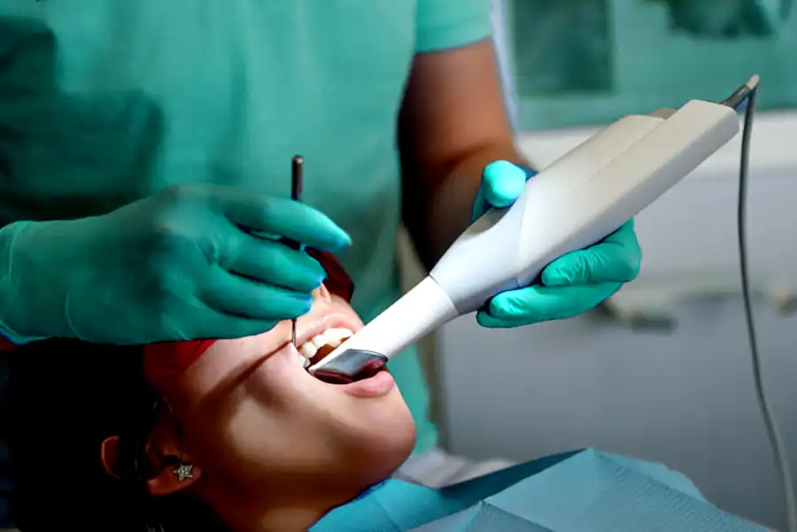
As the dentist or hygienist moves the wand around the inside of your mouth, the device projects a light source—typically structured light or a specialized laser—onto the teeth and gums. High-resolution imaging sensors capture thousands of pictures per second. Specialized software then processes this massive amount of data instantly, stitching the images together to create a perfectly scaled digital replica of your mouth on a computer screen.
The process of learning how to take digital dental impressions is remarkably straightforward for clinical staff. The practitioner simply dries the teeth, gently sweeps the scanning wand over each dental arch, and watches as the digital model builds in real time. There is no waiting for materials to set and absolutely no messy cleanup required afterward.
Traditional Dental Putty vs Digital Scanning
For decades, alginate and PVS (polyvinyl siloxane) putty were the gold standard for taking dental molds. However, when evaluating traditional dental putty vs digital scanning, the digital approach emerges as the clear winner in almost every measurable category.
One of the most celebrated triumphs of this new technology is eliminating gag reflex in dental impressions. For many patients, sitting with a mouth full of thick, expanding putty for up to five minutes is highly anxiety-inducing. The digital wand is non-invasive, breathes easily, and the scanning process can be paused at any time if the patient needs a quick break. This dramatically elevates the overall patient experience with optical impression systems.
But aside from comfort, are digital impressions more accurate than molds? The short answer is a resounding yes. Physical molds are subject to a variety of human errors and material flaws, including shrinkage, tearing, and trapped air bubbles. Digital scans provide a flawless, 1:1 representation of the oral cavity that never distorts over time.
The Core Benefits of a Digital Dentistry Workflow
The transition from physical to digital tools isn’t just about avoiding a messy procedure; it radically improves clinic efficiency. The digital impression system benefits ripple through every phase of a patient’s treatment plan.
1. Unmatched Speed and Efficiency
The advantages of digital dentistry workflow become most obvious when looking at turnaround times. Instead of packaging and mailing fragile physical molds to a dental laboratory—which can take days—digital files are sent via a secure portal in mere seconds. This streamlined communication plays a massive role in reducing chair time for dental crowns, veneers, and implant restorations. Faster lab communication means fewer appointments and quicker final results for the patient.
2. Improved Case Acceptance and Education
Additionally, these devices offer real-time 3D visualization for patients. Seeing a highly detailed, magnified, full-color model of their own teeth on a monitor helps patients better understand their diagnosis. When a dentist can point directly to a cracked tooth or severe wear on a digital 3D model, patients feel more informed and confident in agreeing to proposed treatment plans.
Expanding Applications in Restorative and Orthodontic Care
Achieving high precision 3D oral imaging forms the foundation of several other advanced dental treatments. The data captured by these wands does much more than just replace a physical mold.
In the realm of restorative work, this highly accurate imaging seamlessly integrates with CAD/CAM technology in restorative dentistry (Computer-Aided Design and Computer-Aided Manufacturing). Dentists can digitally design a custom crown or porcelain veneer on their computer and send that design to an in-office milling machine. This workflow allows clinics to offer same-day restorations, completely eliminating the need for temporary crowns.
Orthodontics has experienced a similar revolution. Clinics can now easily export the scanned data as STL files for orthodontic clear aligners. These universal 3D file formats are sent directly to specialized laboratories or companies that manufacture invisible aligners (like Invisalign). The precise fit of these aligners—born from a perfect digital scan—ensures that teeth shift safely and predictably.
Choosing the Right Equipment: What Dentists Need to Know
For practitioners making the leap into the digital age, selecting the right scanner is a critical business decision. If you need a quick guide to modern chairside scanning technology, there are a few key factors to prioritize:
- Ergonomics and Weight: When comparing handheld intraoral imaging devices, ergonomic design is paramount. A lighter, well-balanced wand reduces wrist fatigue for the operator and fits more comfortably into a patient’s mouth, especially for individuals with a restricted jaw opening.
- Software Intelligence: Modern scanners rely heavily on Artificial Intelligence. The best systems feature AI-driven tissue removal, which automatically edits out wandering tongues, fingers, or cheeks from the final digital model in real time, making scanning incredibly fast.
- Open vs. Closed Systems: Opting for an “open” system allows you to easily share files with any laboratory or software platform, offering maximum flexibility for your practice’s future growth.
Final Thoughts
The era of uncomfortable, panic-inducing dental molds is rapidly coming to an end. Dental intraoral scanners represent a monumental leap forward, merging clinical precision with unparalleled patient comfort. By embracing this technology, dental practices can provide faster, more accurate diagnoses while ensuring their patients spend less time in the chair and more time showing off their healthy smiles. Whether you are a patient seeking a modern clinic for your next procedure or a dentist outfitting a new practice, the shift toward digital impressions is a welcome advancement for the entire dental community.
https://dentistbaytowntx.com/dental-care/general-dentistry/digital-x-rays/
Brian L. Porter, DDS
1109 E. James Ave.
Baytown, TX 77520
281-422-3415
View our Google Business Listing